ALIF
| Attending | Vendor | Localization | C arm | Neurophys | Discectomy | Graft height | Graft width | Graft depth | Packing | Pilot holes |
|---|---|---|---|---|---|---|---|---|---|---|
| Miele | Stryker | Flat plate once after exposure only | No | None | End plate shavers and pituitary only | Depends on fit of trial | Always Stryker "small" | Always 25 mm | iFactor | Angled drill |
Preop
You will need a Foley to decompress the bladder.
You may or may not need C arm depending on attending preference. If you use a C arm you will need lead and a C armor.
Measure the preoperative disk angle to give you a sense for the degree of lordosis of the future graft.

Indication
Best for L5/S1, sometimes applicable to L4/5 depending on the location of the bifurcation of the descending aorta into the common iliacs. The location of the venous bifurcation is more important than the arterial bifurcation since these are more delicate structures and more difficult to mobilize. In the image below, the L4/5 disk space is above the bifurcation and this space was not accessible during surgery.

Pay attention to the angle of the L5/S1 disk relative to the pubic symphysis. This will dictate whether you have the angle to approach the disk space.

Positioning
Supine with arms at right angles to the body on arm boards. Red boxes demonstrate where to attach the brackets for the retractor.

Use of a bump is according to surgeon preference. If the approach surgeon uses a bump, place it transversely beneath the pelvis such that the top of the bump is at the top of the iliac crests.
Incision
The incision is variable and depends on approach surgeon's preference. Mark midline between the umbilicus and the pubic symphysis. The images below demonstrate the vertical (right) versus transverse (left) incisions. The circular retractor is attached to the brackets.


Exposure
Done by the abdominal surgeon. They will call when they are ready.
Skin incision is made with a #10 scalpel blade followed by Bovie through the fat to the fascia of the abdominal musculature. The abdominal musculature is opened with the Bovie with a vertical incision in the midline, essentially at the linea alba. Go through the muscle to the preperitoneal space but do not open the peritoneum. Place the general surgery sleeve retractor

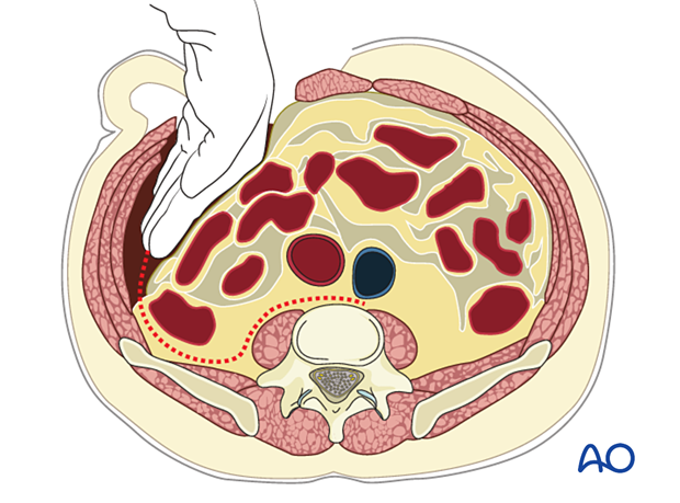
With your hands, navigate the preperitoneal plane laterally and pull the peritoneum with its contents over laterally. Secure in place with the retractors attached to the ring.

Localization
Place a radiopaque instrument (like a Penfield 4) into the candidate disc space and localize on a lateral shot.
Procedure
The neurosurgery team usually scrubs in after the correct level has been verified and the exposure surgeons are happy with the exposure.
When you scrub in, the first thing you should do is to understand from the abdominal surgeon the location of delicate structures such as veins tucked under retractors.
Complete the annulotomy with the long-handle 15-blade.
Specifics of disc removal will depend on the attending. The goal is to remove the disc and cartilaginous end plates without breaking into the bony endplates. There is usually a "fringe" of disc material outside the disc space that does not need to be chased.
- Some prefer to remove disk material with Kerrison and pituitary rongeurs followed by removal of cartilaginous end plate with curettes.
- Others, like Dr. Miele, will go from annulotomy directly to end plate shavers then remove disc fragments with the pituitary. He does not use Kerrisons or curettes at all.

Sizing
This depends on the patient's pathology and attending preferences.
Ask for a trial. Dr. Miele always uses the Stryker "small" graft with a depth of 25 mm. He will vary the height of the graft depending on whether he wants to get neuroforaminal decompression but does not usually bother with greater degrees of lordosis. He does not take C arm shots but will instead decide on height based on how readily a trial of a given height went in and how much indirect decompression it provided.
For other attendings, the width of the trial may be determined by the width of visible disk space. The depth and height of the trial may be determined by taking lateral C arm shots. Some attendings will take an AP C arm shot to verify that the graft is centered on the mid-sagittal plane. Note that the degree of lordosis is constrained by the height of the graft. Decreasing the height may constrain you to lesser degrees of lordosis.
Placement
After the graft is opened and packed with the appropriate material (again, attending preference, Miele uses iFactor), you will have to choose whether to have two holes pointing up or one hole pointing up. For Miele, put the single hole on the side that will be more difficult to screw.
There are two ways to penetrate the bony end plate before placing the screws:
- Use the angled awl with the mallet to create the holes. This can be associated with vertebral body fracture.

- Or use the angled drill. This is the way Miele does it.
Once the screws are placed, turn the locking bolts to keep the screws from backing out.
The abdominal surgeon will close.
